
Within the subdued atmosphere of hospital MRI departments, a groundbreaking transformation is emerging in how researchers detect frontotemporal dementia (FTD), a severe neurological disorder commonly affecting middle-aged adults. Historically challenging to identify until clear symptoms manifest, scientists now are on the brink of predicting the condition far earlier.
An international collaboration spearheaded by Sweden’s Karolinska Institutet has provided strong evidence that minute alterations in the brain’s microscopic structure can be uncovered well ahead of any visible symptoms. This approach moves beyond conventional indicators like brain volume loss or cognitive decline, focusing instead on the movement of water molecules within the brain’s grey matter.
Termed cortical mean diffusivity (cMD), this advanced MRI technique detects micro-level cortical damage that traditional imaging techniques often miss. Remarkably, in individuals genetically susceptible to FTD, these subtle brain changes appeared years prior to clinical diagnosis.

For families with inherited risk, this advance means more than just early detection—it offers precious time for preparation, potential intervention, and possibly altering the disease’s trajectory.
Detecting Hidden Brain Changes
Published recently in Molecular Psychiatry, the study analyzed MRI data from 710 participants, including both mutation carriers and controls. Participants were part of the GENFI project, a global initiative studying genetic forms of FTD.
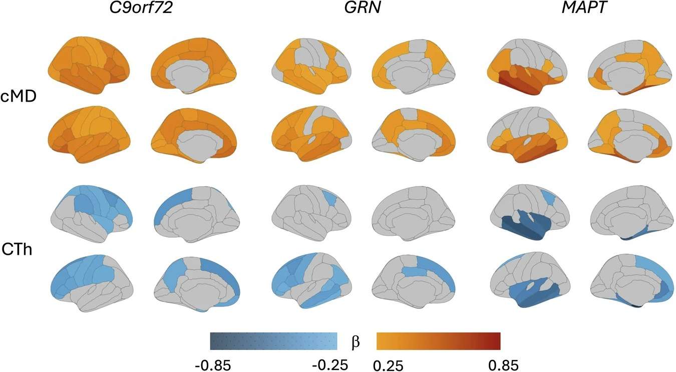
Results were striking: cMD shifts were identified in presymptomatic carriers, individuals harboring genetic mutations but displaying no cognitive symptoms. Unlike standard markers such as cortical thinning, these microstructural changes appeared well in advance. Notably, carriers of the C9orf72 mutation exhibited these changes even with perfectly normal cognitive assessments.
Dr. Elena Rodriguez Vieitez, a prominent neuroimaging expert at Karolinska and study co-author, described this insight as "a glimpse into the silent phase of neurodegeneration." Essentially, this means that medical professionals may eventually identify dementia before individuals recognize any symptoms themselves.
Individualized Risk Profiles
Distinct genetic mutations exhibited varying cMD alteration patterns. For instance, people with MAPT mutations showed early microstructural deterioration during mild symptomatic stages, whereas those with GRN mutations only demonstrated notable changes at more advanced phases.
Understanding these differences allows neurologists to customize monitoring and treatment plans reflecting a person's specific genetic predispositions. It also emphasizes the variability in disease onset and progression among at-risk groups.
“Gaining deeper insight into these patterns,” noted Dr. Caroline Graff, the study’s lead author and head of Neurogeriatrics at Karolinska Institutet, “enhances our ability to support families and design clinical trials with optimal timing.”

Advancing Clinical Trials and Beyond
A critical challenge in neurodegenerative disease research is recruiting suitable candidates for preventative therapy trials, particularly those in the earliest, preclinical stages. Therapeutics are less effective once significant neural damage has occurred.
Utilizing cMD as a biomarker provides a dynamic indicator of disease activity before symptoms manifest, enabling trials to focus on individuals biologically on the path to dementia while still fully functional.
Moreover, this technique has implications beyond FTD. Elevated cMD levels have also been observed in Alzheimer’s disease and ALS, suggesting diffusion MRI’s broader utility in neurodegenerative diagnostics. These findings align with reports from Tech Eye Life Sciences, highlighting the increasing significance of brain microstructure analysis in early disease detection.
A Fundamental Shift in Dementia Research
This work exemplifies a growing emphasis on identifying biological markers rather than relying solely on overt symptoms, aiming for earlier and more precise dementia diagnosis. Techniques such as plasma biomarker analysis and PET scans complement these efforts, all targeting early disease interception and prevention.
While diffusion MRI has been used before, applying it specifically to grey matter at this scale is unprecedented. “We’re moving beyond looking for atrophy,” Rodriguez Vieitez explained. “Our focus is detecting the subtle, initial damage invisible to previous methods.”
Currently suited primarily for research, ongoing refinement may soon enable this method to revolutionize the diagnosis and management of inherited dementias—potentially identifying risk many years before symptoms like memory loss or behavioral changes appear.
- Categories:
- Science


0 comments
Sign in to Comment