
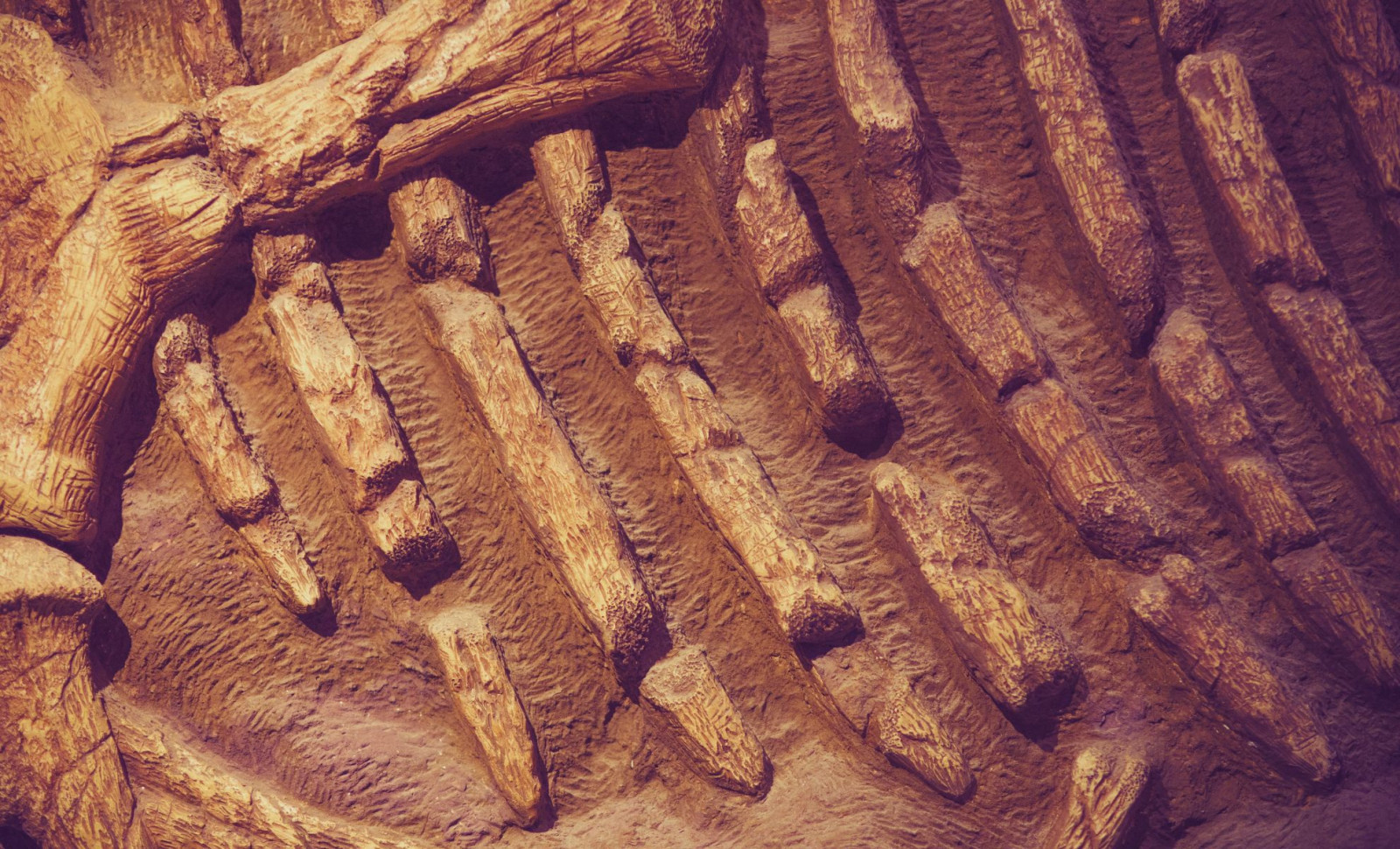
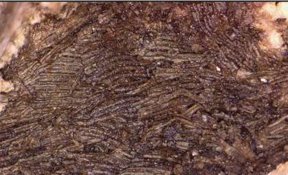
Recent research reveals chemical signatures indicating that traces of hemoglobin, the oxygen-transporting molecule in blood, may be preserved within some dinosaur fossils. Utilizing an advanced spectroscopy method, scientists identified molecular patterns within fossilized structures resembling blood vessels in Tyrannosaurus rex and Brachylophosaurus canadensis specimens.
This discovery contributes fresh insights to a long-standing scientific discussion over whether fragile biological materials can endure for tens of millions of years within fossils.
Employing Laser Spectroscopy to Track Hemoglobin Residues
To delve deeper into the fossilized vessels, Mary Schweitzer, a paleontology expert at North Carolina State University, teamed up with physicist Hans Hallen, an authority on Raman spectroscopy. This technique characterizes chemical substances by interpreting laser light scattering. Their findings, published in a study in Proceedings of the Royal Society A: Mathematical, Physical and Engineering Sciences, employed resonance Raman spectroscopy, which enhances signals from specific molecules.

Fossil samples commonly feature a blend of degraded organic compounds and minerals, presenting challenges for identifying molecules precisely.
“Raman spectroscopy essentially uses light waves to identify a molecule’s energetic ‘fingerprint,'” explained Hallen. He added that “Resonance Raman, which we use here, takes that process one step further by using light that is already tuned to the molecule of interest—so only that type of molecule will resonate.”
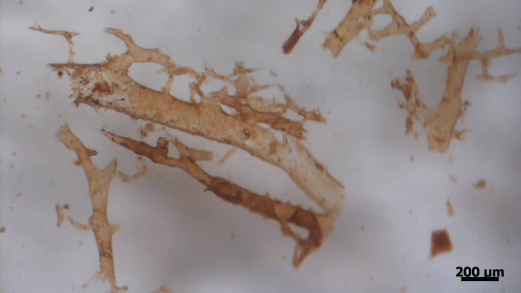
The team concentrated on detecting hemoglobin, which incorporates iron-containing heme rings bound to globin proteins. Identifying these elements would suggest preservation of blood-associated molecular fragments in the vessels.
Hemoglobin Signals Emerge from Laser Analysis
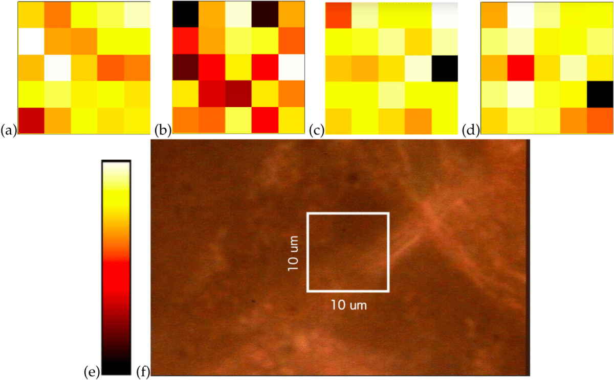
By shining a 532-nanometer green laser on the fossils, researchers observed spectral peaks matching heme groups still linked to globin proteins. These signals were found in vessel-shaped structures from both Tyrannosaurus rex and Brachylophosaurus canadensis fossils.
“The signal increase shows that hemoglobin is present, but changes in the signal also allow us to see that as the hemoglobin degrades, goethite may form on the iron within hemoglobin,” Hallen said in an university report. “We can also pinpoint where the ring-like structure of heme is being damaged.”
To confirm their findings, a second test using a 473-nanometer blue laser was performed. This wavelength is more responsive to heme not bound to proteins, but it produced little signal, supporting the conclusion that the detected molecules are hemoglobin fragments rather than isolated heme or microbial remnants.
The spectral patterns additionally reveal that the hemoglobin is partially broken down, showing chemical modifications expected over such immense preservation timescales.
Iron-Based Chemistry Could Explain Fossil Preservation
The investigation also shed light on mechanisms allowing molecular survival for millions of years. One spectral peak suggests lighter regions within the mineralized vessels correspond to goethite, which, as Hallen explains, “is a biologically linked mineral crystal formed through natural biological processes.”
It’s proposed that interaction between oxygen and the iron atom at the center of heme led to formation of these crystalline minerals, creating zones alternating in oxygen concentration inside the vessels.
In oxygen-poor environments, iron likely drove cycles of oxidation and reduction, causing proteins to cross-link and harden the surrounding tissue. Schweitzer suggests that hemoglobin may have played an active role in this preservation process.
“Heme has been identified in sediments that are much, much older than dinosaurs, so we know that it persists,” she explained. “Understanding why hemoglobin preserves, and the role that heme plays in the process, is really important if we want to know how these ancient molecules survive through time.”
- Categories:
- News


0 comments
Sign in to Comment