
Researchers have successfully examined fossilized embryos of a dinosaur species dating back 200 million years. Using cutting-edge particle accelerator technology, they generated finely detailed 3D reconstructions of the embryos’ cranial structures.
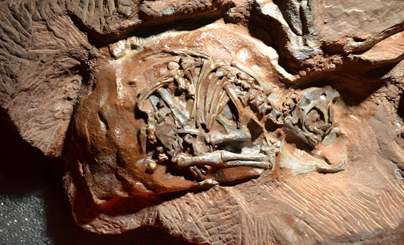
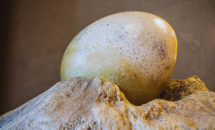
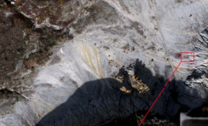
Discovered in 1976 at South Africa’s Golden Gate Highlands National Park, these six fossilized eggs belong to the herbivorous biped Massospondylus carinatus, which thrived during the Early Jurassic era.
Although these embryos were about two-thirds through their development, they provided invaluable insights into dinosaur growth stages, an area previously limited by the delicate nature and small size of such specimens.

Cutting-Edge Imaging Sheds New Light
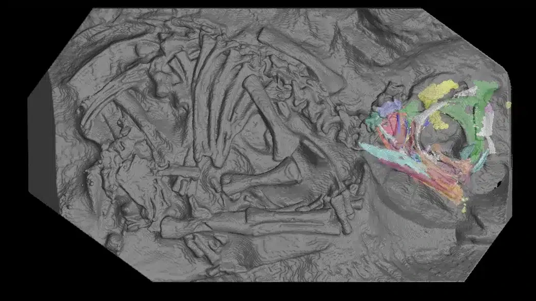
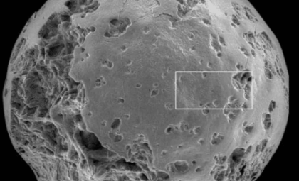
Kimberley Chapelle, a paleontologist at the University of the Witwatersrand, noted that the key advancement occurred when the embryos were scanned at the European Synchrotron Radiation Facility (ESRF) in Grenoble, France. By leveraging ESRF’s intense synchrotron X-rays—much brighter than conventional hospital imaging—they captured images revealing individual bone cells. She stated:
“A synchrotron source is one hundred billion times brighter than a hospital X-ray source,” this made it possible to separate the bones from the surrounding rock and create 3D images of the embryos’ skulls.
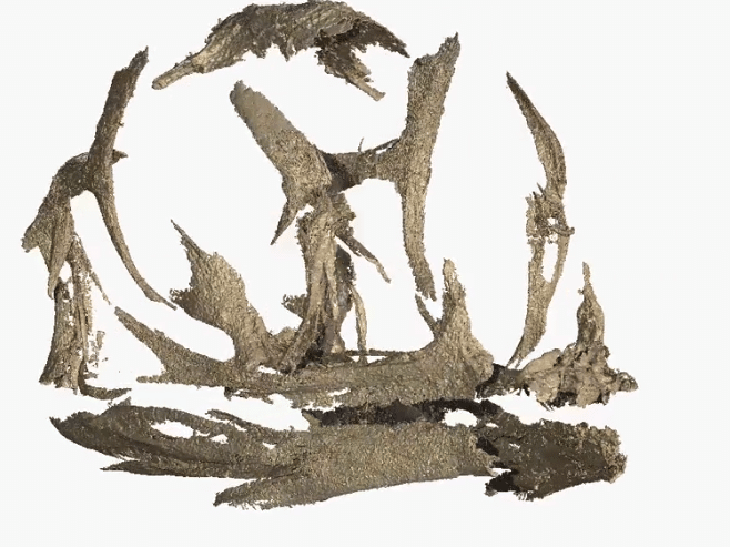
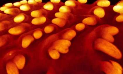
This imaging delivered remarkable clarity, allowing examination of the embryos’ dental and skull features with unprecedented precision. The team identified two varieties of teeth.
“I was really surprised to find that these embryos not only had teeth, but had two types of teeth,” she explained. “The teeth are so tiny; they range from 0.4 to 0.7 mm wide. That’s smaller than the tip of a toothpick.”
Key Insights From the Embryonic Teeth
Finding dual sets of teeth within the Massospondylus embryos surprised the scientific community. One set consists of triangular-shaped teeth likely discarded before hatching, while the other matches the permanent teeth emerging after birth. This two-tiered dental arrangement is reminiscent of modern reptiles such as crocodiles and geckos. Researchers emphasize this similarity in tooth development corresponds closely to patterns observed in extant reptiles.
The research, published in Palaeontology, confirmed similarities between the embryos’ skull formation and that of modern-day reptiles like crocodilians, lizards, and birds. Jonah Choiniere, a Professor of Comparative Palaeobiology, remarked:
“It’s incredible that in more than 250 million years of reptile evolution, the way the skull develops in the egg remains more or less the same.” he added, “Goes to show, you don’t mess with a good thing.”
Tracing Dinosaur Development Through Time
These fossilized eggs rank among the oldest dinosaur embryos ever examined, offering a rare glimpse into the early stages of dinosaur biology. Though small, the Massospondylus embryos illuminate how these ancient creatures formed their skulls and limbs before hatching.
While adults grew to about 16 feet long, these embryos provide a snapshot of their prenatal development. Scientists aim to apply synchrotron imaging to other dinosaur embryos, seeking to uncover additional evolutionary secrets.
Vincent Fernandez, co-author of the study, emphasized the significance of using major facilities such as ESRF, stating that “we can unlock the hidden potential of our most exciting fossils” through this approach.
- Categories:
- Science


0 comments
Sign in to Comment