
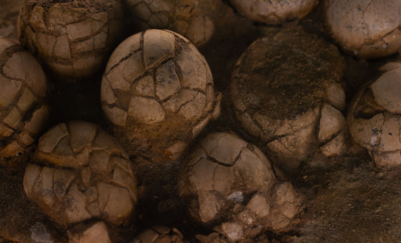
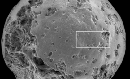
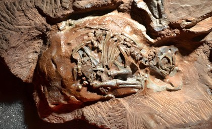
A set of fossilized dinosaur eggs, excavated nearly fifty years ago, has divulged new information through scans performed at a colossal particle accelerator. This advanced imaging enabled scientists to reconstruct the skulls of embryos that have remained intact for 200 million years.
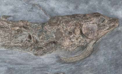
The eggs were originally found in 1976 within South Africa’s Golden Gate Highlands National Park. The cluster includes six eggs housing embryos of Massospondylus carinatus, an early Jurassic bipedal herbivore that could grow up to 16 feet long as an adult.
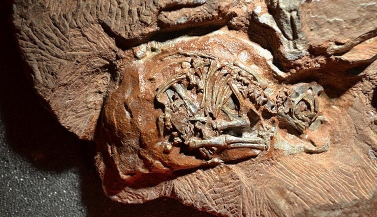
For many years, analyzing these embryos was challenging because of their fragile state and tiny size. Traditional imaging provided limited insights until investigators utilized one of Europe’s most advanced research centers.

Powerful Accelerator Reveals Incredible Microscopic Insights
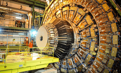
In 2015, scientists Kimberley Chapelle and Jonah Choiniere took the fossils to the European Synchrotron Radiation Facility (ESRF) in Grenoble, France. This facility operates a 2,769-foot circumference ring in which electrons accelerate close to light speed, creating extremely intense X-ray beams.
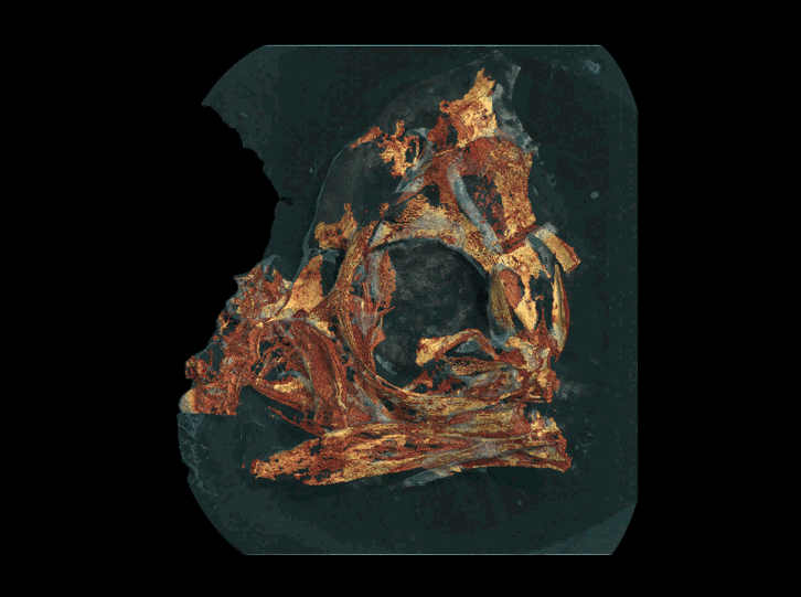
The research, published in Scientific Reports, explains the ESRF’s synchrotron radiation as being substantially brighter than standard medical X-ray machines and far more capable of detecting subtle density variations. This made it possible to clearly distinguish fossilized bone from the surrounding rock matrix.
Chapelle highlighted that standard lab CT scanners cannot achieve such detail. Co-author Vincent Fernandez from the Natural History Museum in London noted only advanced facilities like ESRF could uncover such hidden anatomical features. The scans were so precise that single bone cells became discernible. The data processing spanned nearly three years at the university lab.
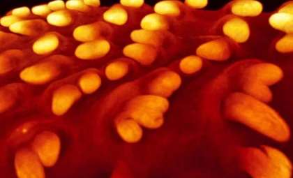
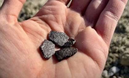
“I was really surprised to find that these embryos not only had teeth, but had two types of teeth. The teeth are so tiny; they range from 0.4 to 0.7 mm wide. That’s smaller than the tip of a toothpick,” she explained.
Two Different Tooth Sets Found in Tiny Skulls
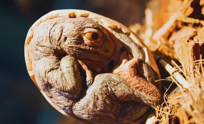
One of the most remarkable findings was that each embryo featured two separate dental patterns. Researchers discovered one set, formed of triangular teeth, likely served as temporary teeth that were shed before hatching. The second set resembled adult teeth, expected to be present when the dinosaur emerged from the egg.
Chapelle expressed surprise at this complexity given the embryos’ tiny scale. The presence of two tooth generations indicates a developed growth process already established during the early Jurassic era.
Fossils Show Development Similar to Modern Reptiles
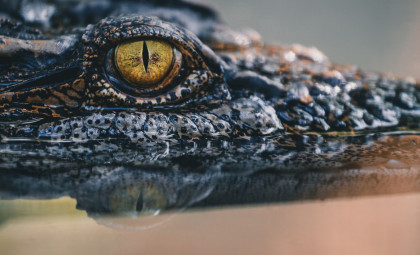
Besides dental features, the scans offered new perspectives on bones formation. Previously scanned limbs from the cluster showed that Massospondylus hatchlings moved on two legs. Scientists identified notable parallels between these embryos and today’s reptiles and birds. As a co-author explained:
“It’s incredible that in more than 250 million years of reptile evolution, the way the skull develops in the egg remains more or less the same. Goes to show—you don’t mess with a good thing.”
Choiniere remarked on how remarkably consistent this reptilian developmental process has been throughout hundreds of millions of years.
- Categories:
- Science


0 comments
Sign in to Comment