

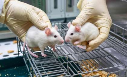
Researchers in Canada placed four mice inside a completely dark enclosure and used highly sensitive cameras to observe them. These cameras detected something invisible to the naked eye: tiny light particles emitted from the living tissues of the animals.
After euthanizing the mice, the team continued recording images. Although the light emissions did not vanish completely, their strength noticeably diminished. This revealed that the light existed predominantly while the animals were alive and faded once they passed.
The study, which appears in a physical chemistry journal, contributes to decades of research on ultraweak biological light emissions. It also draws parallels with early photographic methods often misunderstood as paranormal phenomena before being scientifically explained.

The Subtle Glow of Living Tissue and How It Changes Postmortem
Scientists from the University of Calgary and the National Research Council of Canada reported a marked decrease in visible light emitted by living mice once euthanized, as detailed in The Journal of Physical Chemistry Letters.
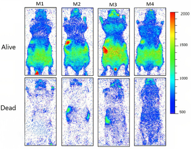
Utilizing electron multiplying charge coupled device (EMCCD) cameras, the researchers captured images of four stationary mice for one hour alive, followed by another hour post-euthanasia. The subjects were kept warm during postmortem imaging to control for temperature effects. The cameras were sensitive enough to detect individual photons within the visible spectrum emitted by cells in both states, noting a statistically significant intensity decrease after death.
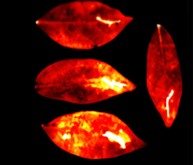
Comparable light emission patterns were observed in thale cress and dwarf umbrella tree leaves when subjected to physical damage or chemical treatments. The scientists observed that damaged areas of leaves consistently emitted more light throughout the 16-hour imaging period.
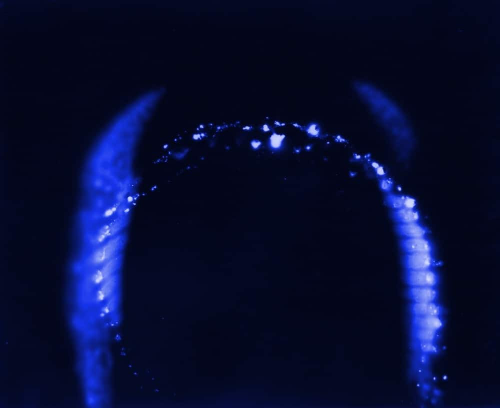
This phenomenon, known as ultraweak photon emission (UPE), has been recognized in isolated biological samples for many years. Previous studies have revealed spontaneous light emissions from cow heart tissue and bacterial colonies, emitting photons across 200 to 1,000 nanometers, as reported in MicrobiologyOpen. The University of Calgary team's work extends these observations to living whole animals.
Electron multiplying CCD cameras, introduced in 2001, boast an on-chip amplification system that detects single photons by multiplying charge signals before readout. This functionality overcomes noise limitations found in traditional CCD or CMOS sensors. According to Oxford Instruments, this technology enables detection of light intensities far weaker than conventional cameras can sense.
The authors suggest reactive oxygen species as the main origin of these emissions. These molecules form in living cells under stress from heat, toxins, infections, or lack of nutrients. Compounds such as hydrogen peroxide promote chemical changes in fats and proteins, which excite electrons to higher states; when electrons fall back, photons are released.
Visible light, ranging from 380 to 700 nanometers on the electromagnetic spectrum, is detected by human eyes through cone cells in the retina. However, the photon flux measured in this study is below the sensitivity of unaided human vision.
The Misinterpretations of Kirlian Photography
This research echoes earlier photo techniques that were mistakenly linked to paranormal effects. Kirlian photography, invented in 1939 by Soviet engineer Semyon Kirlian, captures corona discharges around objects electrically connected to high voltage plates. The method garnered interest in parapsychology in the 1970s, based on the belief that images represented life forces or auras reflecting emotional states.
Scientific scrutiny disproved these claims. Research published in 1973 in the Journal of Applied Physics revealed corona discharge features depend on surface moisture rather than any life energy. Subsequent studies confirmed that cleaning objects removed supposed 'phantom leaf' effects erroneously cited as evidence of lingering energy fields after tissue removal.
The Calgary authors did not employ high voltage or corona discharge techniques. They clarify their research is a biophoton study rather than supporting Kirlian's ideas. As reported by ScienceAlert, they highlight possible uses in noninvasive monitoring of tissue stress in humans, crops, or microbes.
Challenges Before Clinical Adoption
This study's limitations include the small number of mice and the strict lab environment necessary for imaging. Ambient light overwhelms the subtle emissions, requiring complete darkness during data collection. The feasibility of applying this technique for human clinical diagnostics remains to be explored.
The researchers noted that although emissions persisted after death, they were significantly weaker, implying that ongoing metabolic activity is essential for photon generation. Since reactive oxygen species production halts when cellular respiration ends, this aligns with the observed postmortem decline in light intensity.
Neither the National Research Council of Canada nor the University of Calgary has announced further studies or clinical trials to date. Their publication suggests that remote detection of stress via ultraweak photon emission could become a potent, noninvasive tool for medical or agricultural diagnostics but does not provide a timetable for such developments.
- Categories:
- Science


0 comments
Sign in to Comment